
-
Laser
-
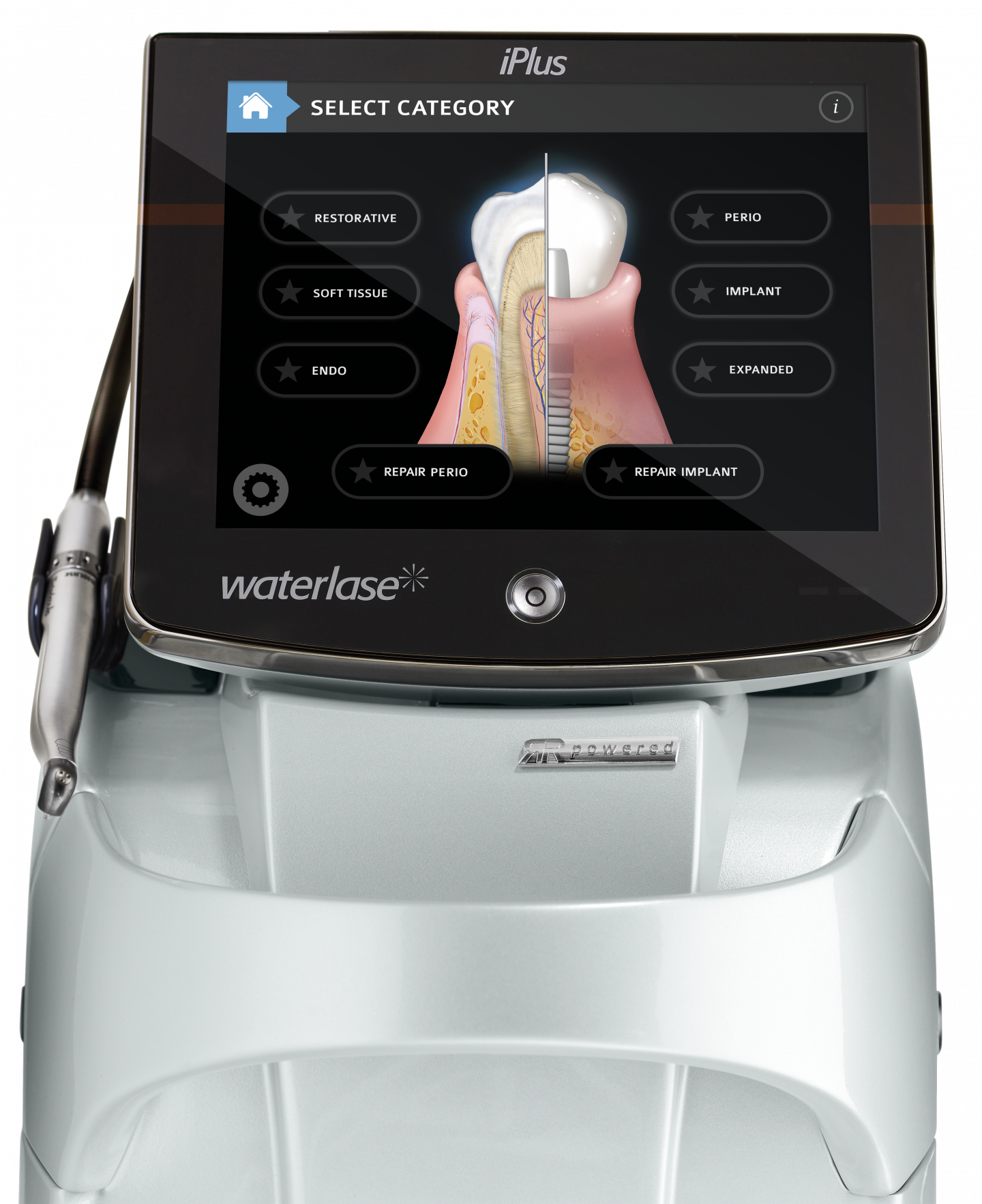
Waterlase iPlusTM - intuitiv und vielseitig
Der Waterlase iPlusTM ist der vielseitigste Dental-Laser auf dem Markt. Dank der einzigartigen Lasertechnologie eignet er sich zur Behandlung von Weich- und Hartgewebe, einschließlich Knochen.
-
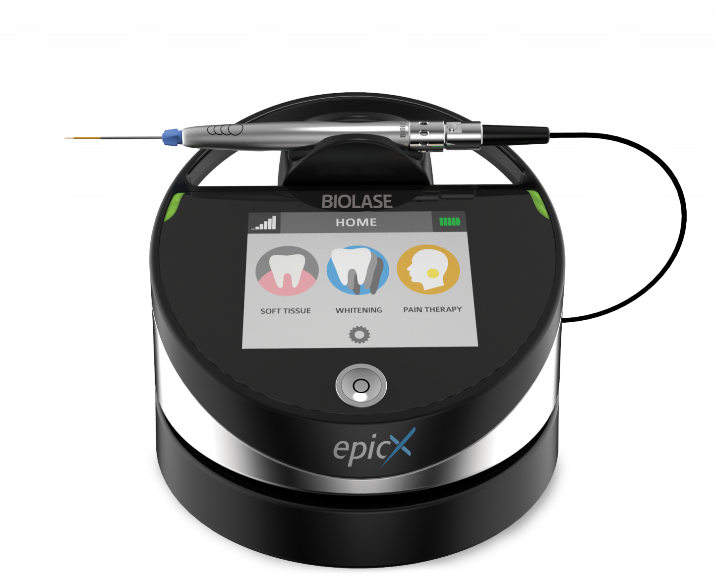
Epic XTM Diodenlaser
Mit dem kleinen, tragbaren Epic XTM Dentallaser schneiden Sie Weichgewebe schonend, schnell und blutungsarm; setzen ihn zur Desinfektion in der Endodontie und Parodontologie ein, stoppen Blutungen, entfernen Hämangiome, lindern Schmerzen, helfen bei Aphten und Herpes und beschleunigen die Bleaching Behandlung.
-
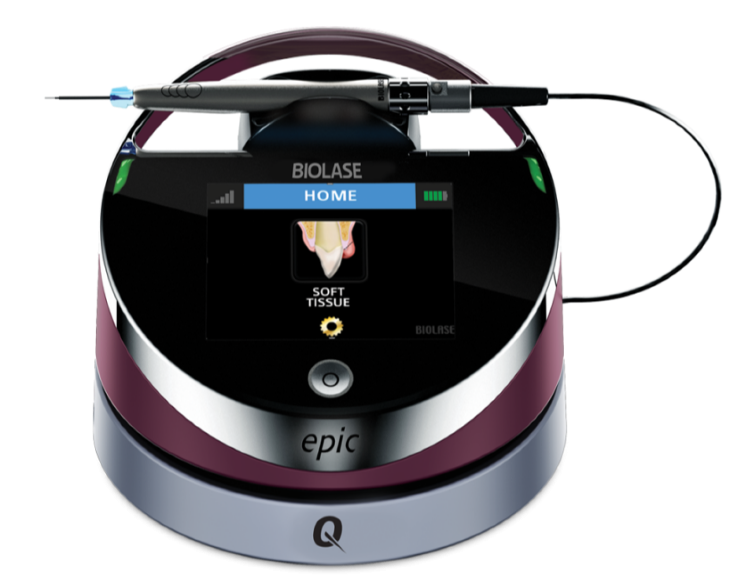
Epic QTM Diodenlaser
Der Epic QTM vereint Langlebigkeit mit Zuverlässigkeit und bietet die neueste
Technologie zur Behandlung von Weichgewebe
in einem tragbaren, erschwinglichen Paket.
-
-
Events
-
BIOLASE Veranstaltungen
In unserem Veranstaltungskalender finden Sie alle nationalen und internationalen Messen, Kongresse, Workshops und Webinare auf einen Blick – wir freuen uns auf Sie!
-
BIOLASE Webinararchiv
Hier können Sie unsere bereits abgehaltenen Webinare jederzeit, nach kostenloser Registrierung abspielen.
Wählen Sie einfach den Fachbereich oder den Referenten für den Sie sich interessieren. -
BIOLASE Webinararchiv international
Hier finden Sie bereits archivierte Webinare unserer internationalen Referenten. Die Webinare können jederzeit abgespielt werden.
zum Archiv -
BIOLASE Academy
Die BIOLASE Academy ist unsere zusätzliche Plattform für internationale Webinare, die jederzeit nach Bedarf abgespielt werden können.
zur BIOLASE Academy
-
- Klinische Studien
- Biolase
- Downloads
- Kontakt
- Shop
Klinische Studien zur Oralchirurgie mit dem Dental-Laser
| Groups | Link | Author | Title | Journal | Year | Rating |
|---|---|---|---|---|---|---|
| Soft Tissue | DOI URL | Arora, S., Lamba, A.K., Faraz, F., Tandon, S. and Chawla, K. | Treatment of Peripheral Ossifying Fibroma using Er,Cr:YSGG Laser : Report of Two Cases [Abstract] |
International Journal of Laser Dentistry Vol. 3(3), pp. 94-99 |
2013 | rank4 |
| Abstract: Peripheral ossifying fibroma (POF) is a non-neoplastic growth on the gingiva thought to arise from the periodontal ligament. Histopathologic analysis always reveals immature bone and osteoid within the lesion. Surgical excision is the treatment of choice. This report comprises of two cases of POFs which were removed using an Er,Cr:YSGG laser. Two patients of age 36 and 25 years reported with the growths which were causing constant irritation as well as esthetic problem. Lesions were removed using an Er,Cr:YSGG laser. The healing was uneventful and no suture or analgesic was required. The histopathological report confirmed them as POF. No relapse was observed till 1 year after surgery. Therefore with this laser, there is relatively bloodless and painless surgery and in postsurgical course there is minimal swelling and scarring so patient's compliance is much better with this laser as compared to other techniques and lasers. Keywords: | ||||||
| Soft Tissue | DOI URL | Castelo-Baz, P., Seoane-Romero, J.M., García-Caballero, L., Suárez-Peñaranda, J.M., Romero-Méndez, M.A. and Varela-Centelles, P. | Unifocal orofacial granulomatosis in retromolar mucosa: surgical treatment with Er,Cr:YSGG laser. [Abstract] |
Journal of clinical and experimental dentistry Vol. 6(2), pp. e189-92 |
2014 | rank4 |
| Abstract: Orofacial granulomatosis is defined by permanent or recurrent swelling of orofacial tissues with different multiform and multifocal clinical patterns. An 11-year old boy presented with a 2-month history of mucosa enlargement. Intraoral examination revealed an erythematous, polylobulated, exophytic lesion with a smooth surface located in retromolar mucosa, non-tender and non-infiltratated to palpation. The diagnosis was inflammatory lesion compatible with pyogenic granuloma and laser excision was decided. Haematological parameters were within normal range, as well as chest Xrays. These findings lead to a diagnosis of non-symptomatic orofacial granulomatosis, whose early diagnosis can minimize the impact of systemic-related disorders, like Chron's disease. Key words:Laser, orofacial granulomatosis, childhood, oral lesions, diagnosis. | ||||||
| Incision | DOI | Cercadillo-ibarguren, I., España-tost, A., Arnabat-domínguez, J., Valmaseda-Castellon, E., Berini-Aytes, L. and Gay-Escoda, C. | Histologic evaluation of thermal damage produced on soft tissues by CO2, Er,Cr:YSGG and diode lasers [Abstract] |
Medicina Oral, Patologia Oral y Cirugia Bucal Vol. 15(6), pp. 912-918 |
2010 | rank5 |
| Abstract: Objective: The aim of this in vitro experimental study was to perform histological evaluation of the thermal ef- fect produced on soft tissue irradiated with CO2 , Er,Cr:YSGG or diode lasers. Study design: Porcine oral mucosa samples were irradiated with Er,Cr:YSGG laser at 1 W with and without water / air spray, at 2 W with and without water / air spray, and at 4 W with water / air spray, with CO2 laser at 1 W, 2 W, 10 W, 20 W continuous mode and 20 W pulsed mode and diode laser at 2W, 5W, and 10W pulsed mode. The thermal effect was evaluated measuring the width of damaged tissue adjacent to the incision, stained positively for hyalinized tissue with Hematoxylin-Eosin and Masson Trichrome stains. Besides, histological changes in the irradiated tissue were described using subjec- tive grading scales. Results: The evaluated lasers developed a wide range of thermal damage with significant differences between groups. The samples with lowest thermal effect were those irradiated with Er,Cr:YSGG laser using water / air spray, followed by CO2 and diode lasers. Conclusions: Emission parameters of each laser system may influence the thermal damage inflicted on the soft tissue, however, the wave length of each laser determines the absorption rate characteristics of every tissue and the thermal effect. | ||||||
| Soft Tissue | URL | Gholami, L., Moghaddam, S.A., Ayoub, M., Ladiz, R., Manesh, Z.M., Hashemzehi, H., Fallah, A. and Gutknecht, N. | Comparison of gingival depigmentation with Er , Cr : YSGG laser and surgical stripping , a 12-month follow-up [Abstract] |
Lasers in Medical Science Vol. April, pp. 1-10 |
2018 | rank5 |
| Abstract: Gingival melanin hyperpigmentation is an esthetic concern for many individuals. In this study, we compared the standard surgical removal method with two different Er,Cr:YSGG laser settings in order to find the best treatment method. In 33 dental arches, the following three treatment groupswere comparatively evaluated: (1) surgical stripping, (2) removalwith laser setting 1 (4.5W, 50Hz, 100% water, 80% air, 60 μs, 800 μm Tip;MZ8), and (3) laser setting 2 (2.5W, 50 Hz, 20% water, 40% air, 700 μs, 800 μmTip; MZ8).We comparatively evaluated pain, patient satisfaction and wound healing, treatment time, and the amount of bleeding. Re- pigmentation was evaluated after 1 and 12 months by Hedin and Dummet pigmentation scores. Laser setting 1 had the best results regarding pain and patient satisfaction, although not statistically significant (P> 0.05).Wound healing results were better using lasers compared to surgical stripping (P< 0.05). Laser setting 1 was a faster procedurewithmild amounts of bleeding. The least amount of bleedingwas seen with laser setting 2. After 1month, only twocases of the laser setting 2-treated areas showed an isolated pigmented area in the papilla; at 12months, the mean Hedin indexes were still less than 2 and mean Dummett index less than 1 in all treatment techniques, with the lowest scores seen in the laser setting 1 sites. Based on our results, Er,Cr:YSGG laser can be more convenient for gingival depigmentation compared to surgical blade. Although not statistically significant, laser setting 1with shorter pulse duration and higher water spray showed better overall results. However, laser setting 2, with longer pulse duration and less water spray, resulted in better coagulative effects and can be used to control bleeding wherever necessary in clinical practice. | ||||||
| Soft Tissue | DOI URL | González-Mosquera, a., Seoane, J., García-Caballero, L., López-Jornet, P., García-Caballero, T. and Varela-Centelles, P. | Er,CR:YSGG lasers induce fewer dysplastic-like epithelial artefacts than CO(2) lasers: an in vivo experimental study on oral mucosa. [Abstract] |
The British journal of oral & maxillofacial surgery, pp. 5-9 | 2011 | rank5 |
| Abstract: Our aim was to assess wounds made by lasers (CO(2) and Er,Cr:YSGG) for their epithelial architectural changes and width of damage. We allocated 60 Sprague-Dawley(®) rats into groups: glossectomy by CO(2) laser at 3 different wattages (n=10 in each); glossectomy by Er,Cr:YSGG laser at two different emissions (n=10 in each), and a control group (n=10). Histological examination assessed both prevalence and site of thermal artefacts for each group. Both lasers (CO(2) and Er,Cr:YSGG) caused the same type of cytological artefacts. The 3W Er,Cr:YSGG laser produced the fewest cytological artefacts/specimen, and was significantly different from the other experimental groups: 3W CO(2) laser (95% CI=0.8 to 1.0); the 6W CO(2) laser (95% CI=0.1 to 2.0) and the 10W CO(2) laser (95% CI=1.1 to 3.0). CO(2) lasers (3-10W) generate epithelial damage that can simulate dysplastic changes with cytological atypia that affects mainly the basal and suprabasal layers. Irradiation with Er,CR:YSGG laser (2-4W) produces significantly fewer cellular artefacts and less epithelial damage, which may be potentially useful for biopsy of oral mucosa. | ||||||
| Crown Troughing | URL | Jetter, C. | Soft-Tissue Management Using an Er, Cr:YSGG Laser During Restorative Procedures [Abstract] |
Dentistry India Vol. 2(5) |
2008 | rank3 |
| Abstract: Effective management of gingival tissues in restorative dentistry poses a challenge to practitioners. Many methods and materials are available to dental professionals to manage tissue. This article will demonstrate the use of an Er,Cr:YSGG laser as an effective, minimally invasive technology to manage soft tissue during restorative procedures. | ||||||
| Incision | URL | Jin, J., Lee, S. and Yoon, H. | A comparative study of wound healing following incision with a scalpel, diode laser, or Er,Cr:YSGG laser [Abstract] |
Acta Odontologica Scandinavica Vol. 68(4), pp. 5 |
2010 | rank4 |
| Abstract: OBJECTIVE:
This study compared wound healing following incisions with either a scalpel, a diode laser or an Er,Cr:YSGG laser in guinea pig oral mucosa. MATERIAL AND METHODS: Three types of wound were made randomly with either a stainless-steel scalpel, a diode laser or an Er,Cr:YSGG laser in the buccal mucosa of 24 guinea pigs. Five guinea pigs were sacrificed on each of Days 1, 3, 5 and 7 post-surgery. Four guinea pigs were sacrificed on Day 14 post-surgery. Biopsy samples from each oral mucosa wound were examined using light microscopy and the expression of tumor necrosis factor (TNF)-alpha and transforming growth factor (TGF)-beta1 was determined by immunohistochemical staining. The expression of TNF-alpha and TGF-beta1 was evaluated by calculating the percentage of positively stained cells and immunostaining intensity was evaluated using a scale ranging from 0 to 3. RESULTS: Infiltration of inflammatory cells decreased rapidly at Day 5 post-surgery in all three groups of animals. The highest level of TNF-alpha expression was found at Day 1 post-surgery for the diode laser wounds. The intensity of TNF-alpha immunostaining was highest at Day 3 post-surgery and lowest at Day 7 post-surgery for all three groups of animals. For the scalpel wounds, a lower level of TGF-beta1 expression was seen until Day 3 post-surgery and a higher level from Day 7 post-surgery compared to laser wounds. The intensity of TGF-beta1 immunostaining was highest at Day 1 post-surgery for the diode laser wounds. CONCLUSIONS: The diode laser is considered a good cutting device for oral mucosa; however, more tissue damage occurs than with the use of a scalpel or an Er,Cr:YSGG laser. Larger studies will be needed before fully endorsing the widespread use of the diode laser. |
||||||
| Frenectomy, Microstructure / Mineral Content | URL | Olivi, G., Chaumanet, G., Genovese, M.D., Beneduce, C. and Andreana, S. | Er,Cr:YSGG laser labial frenectomy: A clinical retrospective evaluation of 156 consecutive cases [Abstract] |
General Dentistry | 2010 | rank4 |
| Abstract: The labial frenum may impede oral hygiene and result in diastema between anterior teeth and traction of the attached gingiva. Surgical removal of the frenum during puberty has been recommended for these patients. This article clinically evaluates the efficacy of an Er,Cr:YSGG laser in removing the labial frenum in an adolescent and pre-pubescent population. Using an Er,Cr:YSGG laser at a power setting of 1.5 W or less and 20–30 pulses per second, a total of 156 frenectomies were performed on 143 children. Patients returned for recall visits at 3, 7, 21, and 30 days and at one, two, and three years. Surgical areas were checked for adverse events, recurrency of frenum, and functional complications. Patient acceptance was also evaluated by using the Wong-Baker FACES pain rating scale. Thirteen recurrences were reported in the adolescent population at 21 or 30 days, all of which required re-intervention; however, only two cases displayed recurrence of the frenum. None of the three pre-pubescent cases required additional intervention, maintaining acceptable clinical results after three years. Patient acceptance was very high, and no postoperative adverse events were reported. Received: | ||||||
| Frenectomy | DOI | Pié-Sánchez, J., España-Tost, A.J., Arnabat-Domínguez, J. and Gay-Escoda, C. | Comparative study of upper lip frenectomy with the CO2 laser versus the Er,Cr:YSGG laser [Abstract] |
Medicina Oral, Patologia Oral y Cirugia Bucal Vol. 17(2), pp. e228-e232 |
2012 | rank5 |
| Abstract: Objectives: To compare upper lip frenulum reinsertion, bleeding, surgical time and surgical wound healing in frenectomies performed with the CO2 laser versus the Er, Cr:YSGG laser. Study design: A prospective study was carried out on 50 randomized pediatric patients who underwent rhomboidal resection of the upper lip frenulum with either the CO2 laser or the Er,Cr:YSGG laser. Twenty-five patients were assigned to each laser system. All patients were examined at 7, 14, 21 days and 4 months after the operation in order to assess the surgical wound healing. Results: Insertion of the frenulum, which was preoperatively located between the upper central incisors, migrated to the mucogingival junction as a result of using both laser systems in all patients. Only two patients required a single dose of 650 mg of paracetamol, one of either study group. CO2 laser registered improved intraoperative bleeding control results and shorter surgical times. On the other hand, the Er,Cr:YSGG laser achieved faster healing. Conclusions: Upper lip laser frenectomy is a simple technique that results in minimum or no postoperative swelling or pain, and which involves upper lip frenulum reinsertion at the mucogingival junction. The CO2 laser offers a bloodless field and shorter surgical times compared with the Er,Cr:YSGG laser. On the other hand, the Er,Cr:YSGG laser achieved faster wound healing. | ||||||
| Frenectomy | URL | Pradhan, S., Yasmin, E. and Mehta, A. | Management of Posterior Ankyloglossia using the Er,Cr:YSGG Laser [Abstract] |
International Journal of Laser Dentistry Vol. 2(2), pp. 41-46 |
2012 | rank3 |
| Abstract: Ankyloglossia or tongue-tie, refers to a minor anomaly in the attachment of the membrane or frenum that attaches the tongue to the floor of the mouth, which may interfere with normal function and mobility of the tongue. Ankyloglossia is a poorly recognized and inadequately defined condition and has been reported to cause breastfeeding difficulties, dental issues and speech problems. This article discusses assessment criterions to diagnose posterior ankylogossia and lays down treatment protocol in neonates using the Er,Cr:YSGG (Waterlase MD). | ||||||
| Incision | URL | Rizoiu, I.M., Eversole, L.R. and Kimmel, A.I. | Effects of an erbium, chromium: yttrium, scandium, gallium, garnet laser on mucocutanous soft tissues [Abstract] |
Oral Surgery Oral Medicine Oral Pathology Vol. 82(4), pp. 386-395 |
1996 | rank4 |
| Abstract: OBJECTIVES:
Lasers are effective tools for soft tissue surgery. The erbium, chromium: yttrium, scandium, gallium, garnet laser is a new system that incorporates an air-water spray. This study evaluates the cutting margins of this laser and compares healing with laser and conventional scalpel and punch biopsy-induced wounds. STUDY DESIGN: New Zealand white rabbits were divided into serial sacrifice groups; the tissues were grossly and microscopically analyzed after laser and convential steel surgical wounding. RESULTS: Wound margins were found to show minimal edge coagulation artifact and were 20 to 40 mm in width. Laser wounds showed minimal to no hemorrhage and re-epithelialization and collagenization were found to occur by day 7 in both laser and conventional groups. CONCLUSIONS: The new laser system is an effective soft tissue surgical device; wound healing is comparable to that associated with surgical steel wounds. The minimal edge artifact observed with this laser system should allow for the procurement of diagnostic biopsy specimens. |
||||||
| Crown Troughing | URL | Scott, A. | Use of an erbium laser in lieu of retraction cord: A modern technique [Abstract] |
General Dentisry, pp. 116-119 | 2005 | |
| Abstract: The two-cord retraction technique is used for troughing around a crown to achieve biologic width. This technique can cause significant discomfort for the patient and offers several potential clinical disadvantages for the dentist, of which unpredictable tissue recession is the most significant. The clinical case presented in this article compares the use of the standard two-cord retraction technique with that of an 2,780 nm erbium-class dental laser to determine which method achieves an accurate, easily readable impression while respecting the biologic width. Using an erbium laser to achieve the trough prior to placing an indirect restoration results in little or no postoperative discomfort for the patient; in addition, the erbium laser reduces intraoperative complications related to tissue recession and patient discomfort while providing consistently accurate impressions. | ||||||
| Fibroma | DOI | Seoane, J., González-Mosquera, A., López-Niño, J., García-Caballero, L., Aliste, C., Seoane-Romero, J.M. and Varela-Centelles, P. | Er,Cr:YSGG laser therapy for oral leukoplakia minimizes thermal artifacts on surgical margins: a pilot study [Abstract] |
Lasers in Medical Science Vol. 28, pp. 1591-1597 |
2013 | rank4 |
| Abstract: Laser use for biopsy of suspicious lesions may simulate cytological atypia at the margin of the incisions, challenging pathological diagnosis. Erbium, chromium: yttrium–scandium–gallium–garnet (Er,Cr:YSGG) laser has shown promising results in experimental models by inducing fewer artifacts. The aims of this study were to examine the thermal wounds induced by Er,Cr:YSGG laser in a short series of oral leukoplakias in terms of cytological and epithelial architectural changes and also to assess the width of the thermal damage lateral to the incision. Four oral leukoplakia patients entered the study and underwent complete surgical excision of their lesions by using Er,Cr:YSGG laser. Patients were weekly controlled until complete healing was accomplished. The patients were included on the existing follow-up program for these lesions thereafter. Study samples were routinely processed by the same technician and double-blindedly studied by two pathologists until a consensus was reached for each case. The pathological analysis of the samples revealed no autolysis and no fixation- or handling-related artifacts. However, cellular and nuclear polymorphism could be observed in two samples. Loss of intercellular adherence was the most frequent thermal artifact in this series; all pseudodysplastic artifacts recognized in the study were of low intensity and located at the basal and suprabasal layers of the leukoplakias’ epithelium. The width of the thermal damage at the edge of the incision scored an average of 26.60 ± 25.3 μm. It is concluded that irradiation with Er,Cr:YSGG laser induces a minimal amount of thermal artifacts at the surgical margins of oral leukoplakias and avoids diagnostic interferences with real dysplastic borders. | ||||||
| Gingivectomy | URL | Soares, F.M., Tarver, E.J., Bimstein, E., Shaddox, L.M. and Bhattacharyya, I. | Gingival Overgrowth in a Child With Arthrogryposis Treated With a Er,Cr:YSGG Laser: A Case Report [Abstract] |
Pediatric Dentistry Vol. 31(01) |
2009 | rank4 |
| Abstract: The present manuscript reports a case of a 21/2 year old girl, diagnosed with arthrogryposis, presenting increasing gingival hyperplasia which was treated with Er,Cr:YSGG Laser. The patient was treated under general anesthesia by the Pediatric Dentistry and Periodontics departments. Er,Cr:YSGG laser G6 tip was used at 1.50 watts, 20 pps, 8% water and 11% air, which is recommended by the manufacturer. Scalpel and periodontal curettes were used to complement the laser. Tissue samples from the anterior maxilla, anterior mandible and palatal sites were formalin-fixed and submitted for evaluation. The samples biopsied revealed prominent hyperplasia of the fibrous connective tissue with areas of the epithelium exhibiting pseudoepitheliomatous hyperplasia. At 1 week and 3 months follow up, oral examination showed appropriate healing of gingival tissue. The use of Er,Cr:YSGG laser in the present case proved to be effective in the removal of large amounts of hyperplasic gingival tissue and resulted in fast heeling and mild discomfort. | ||||||
| Fibroma | URL | Trajtenberg, C. and Abidi, S. | Removal of an irritation fibroma using an Er,Cr:YSGG laser: a case report [Abstract] |
General Dentistry Vol. 56(7) |
2008 | rank3 |
| Abstract: The irritation fibroma is a benign proliferation that occurs as a response to local irritation. It is an elevated pedunculated or sessile lesion that ranges in size from a few millimeters to a few centimeters and is normal in color, although it may appear to be more pale than the normal mucosa. This article describes the case of a 39-year-old woman with an asymptomatic nodular lesion that involved the left buccal mucosa. The lesion was removed using an Er,Cr:YSGG laser. Only topical anesthesia was applied to the fibroma and adjacent mucosa and no infiltration was required. No pain medication or antibiotics were required after surgery and wound healing was excellent and achieved rapidly. The oral pathology report confirmed the presurgical clinical diagnosis. Laser excision is a modern approach for treating oral soft tissue lesions and should be considered as an alternative to conventional scalpel surgery. | ||||||
| Fibroma | URL | Walinski, C. | Irritation fibroma removal: a comparison of two laser wavelengths [Abstract] |
General Dentistry Vol. 52(3) |
2004 | |
| Abstract: Laser wavelengths are available that offer dentists treatment options for both hard and soft oral tissues. This article discusses the benefits of removing hypertrophic soft tissue by using an all-tissue laser. A clinical case is presented involving the use of both an 810 nm diode laser and a 2,780 nm Er,Cr:YSGG laser to remove two irritation fibromas, both located on the left cheek of the same patient. This unique perspective was ideal for comparing the healing of each wound. The Er,Cr:YSGG appeared to have a less traumatic effect on target tissue and offered improved postoperative healing, faster recovery time, and less trauma than traditional surgical modalities. | ||||||
| Gingivectomy | URL | Wang, X., Zhang, C. and Matsumoto, K. | In vivo study of the healing processes that occur in the jaws of rabbits following perforation by an Er,Cr:YSGG laser [Abstract] |
Lasers in Medical Science | 2005 | rank3 |
| Abstract: This study investigates the healing process that takes place in the bone and soft tissue of the maxilla and the mandible after perforation by an Er,Cr:YSGG laser device. The jaws of New Zealand white rabbits were irradiated with an Er,Cr:YSGG laser, forming wounds 0.4 mm in diameter. Irradiation parameters were as follows: repetition rate was 20 pulse/s, pulse duration was 140–200 μs, power was 2 W, exposure time was 10 s, energy density was 80 J/cm2. After sacrifice at 0–56 days post-surgery, gross observations and histological examinations were performed. Effective hemostasis was achieved after Er,Cr:YSGG laser surgery. There was a minimal delay before the healing began. After 56 days all of the bone defects had been completely replaced by new bone. In conclusion, the Er,Cr:YSGG laser allows precise surgical ablation with minimal thermal damage to adjacent tissues in vivo. The overall subsequent healing was favorable. This laser may potentially be used in minor oral surgery. | ||||||
| Soft Tissue | DOI URL | Yilmaz, H.G., Albaba, M.R., Caygur, A., Cengiz, E., Boke-Karacaoglu, F. and Tumer, H. | Treatment of recurrent aphthous stomatitis with Er,Cr:YSGG laser irradiation: A randomized controlled split mouth clinical study [Abstract] |
Journal of Photochemistry and Photobiology B: Biology Vol. 170, pp. 1-5 |
2017 | rank5 |
| Abstract: The present randomized controlled split mouth clinical study aimed to investigate the efficacy of Er,Cr:YSGG laser irradiation on pain reduction and healing rate of recurrent aphthous stomatitis. 40 patients with RAS were recruited for this study and RAS ulcerations of each patient were randomly assigned to the control or test group. In the test group, Er,Cr:YSGG laser with non-contact mode was used to irradiate RAS lesions. In the placebo group, RAS lesions were irradiated with the same device without laser emission. Pain was evaluated with visual analog scale (VAS) while a clinician graded healing of RAS (HRAS). In the placebo group at immediate; scores of VAS presented no statistically significance; in the test group, laser application showed significant pain reducing, at 1st day control. In the test group, a significantly healing effect at 1st day control was observed and this effect was maintained throughout the study. In the placebo group, scores of HRAS were statistically significant at controls on 3, 7, 10??days. Statistically significant difference between the scores of VAS and HRAS was found for all control days except day 10 according to the intergroup comparisons. Based on these findings, Er,Cr:YSGG laser application at 0.25??W without water may be appropriate to reduce pain and also accelerate the healing of RAS. | ||||||
Innovative Dentallaser für Knochen, Hart- und Weichgewebe. BIOLASE ist mit der Dentallaser-Technologie führend in der Zahnmedizin sowie in der Mund-, Kiefer- und Gesichtschirurgie.